Have you ever wondered how you effortlessly lift a heavy object, or sprint across a finish line? The answer lies within the intricate world of skeletal muscle, the powerhouses that drive movement in our bodies. Imagine a symphony of tiny muscle fibers, working in perfect harmony, orchestrated by a complex network of proteins, all under the direction of our nervous system. This intricate dance, happening at the microscopic level, is the foundation of our physical abilities. In this exploration, we delve into the fascinating world of skeletal muscle anatomy and organization, revealing the secrets behind our strength and agility.

Image: anatomyworksheets.com
Understanding the microscopic structure of skeletal muscle unlocks a wealth of knowledge, not only for aspiring athletes and fitness enthusiasts, but also for anyone seeking a deeper understanding of the human body. From the basic unit of a muscle fiber, the sarcomere, to the intricate interplay of proteins like actin and myosin, the structure of skeletal muscle is a testament to the brilliance of nature’s design. This review sheet will serve as your guide, leading you through the labyrinth of muscle tissue, shedding light on its complex organization, and revealing the secrets behind its incredible functionality.
The Building Blocks of Power: Delving into the Muscle Fiber
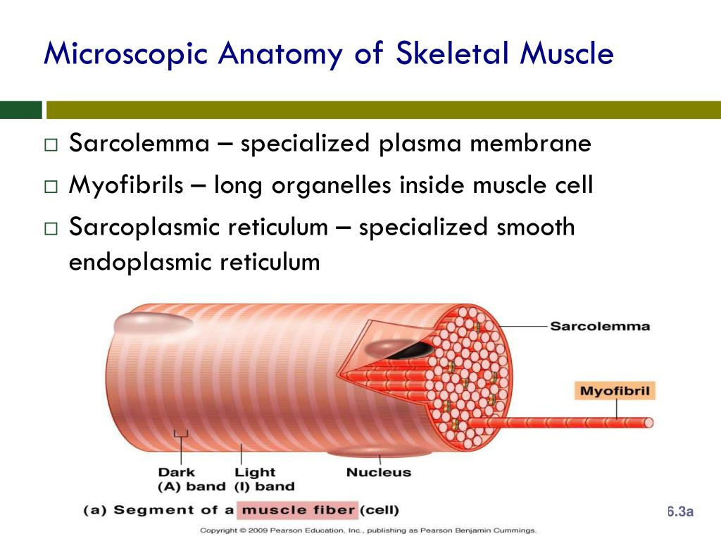
Let’s begin our journey by entering a single muscle fiber, the fundamental unit of skeletal muscle. Each fiber is essentially a long, cylindrical cell, packed with bundles of protein filaments called myofibrils. These myofibrils, like tiny threads woven together, are responsible for the muscle’s ability to contract and generate force. Picture a stack of coins – the myofibrils appear as these stacks, perfectly aligned, and repeating throughout the entire muscle fiber. Each segment of this stack, resembling a single coin in our analogy, is called a sarcomere.
The sarcomere, considered the functional unit of skeletal muscle, is where the magic happens. Inside the sarcomere, we find two primary protein filaments: actin and myosin. Imagine a rope ladder, with actin forming the rungs and myosin the vertical strands. This is a simplified representation of the arrangement of these two key proteins. Think of the myosin filaments as tiny molecular motors, equipped with heads that can bind to the actin filaments. When a signal arrives from the nervous system, the myosin heads latch onto the actin filaments, pulling them closer together. This pulling action, repeated across countless sarcomeres, leads to the shortening of the muscle fiber, resulting in the characteristic muscle contraction.
The Sarcomere: A Closer Look at the Functional Unit
Let’s delve deeper into this fascinating sarcomere. It’s defined by distinct regions, each with its own role in the contraction process. At the edges of the sarcomere, we find the Z-lines, anchoring the thin actin filaments. Think of these Z-lines as the walls holding the rope ladder steady. Spanning the entire length of the sarcomere are the thick myosin filaments, with their heads extending towards the actin filaments. This arrangement mirrors the arrangement of a rope ladder, with the rungs (actin) being pulled closer together by the ropes (myosin).
The region in the middle of the sarcomere, where only myosin filaments are present, is called the H-zone. Visualize this as the central portion of the ladder, where only the ropes are present. During contraction, the actin filaments slide past the myosin filaments, pulling the Z-lines closer together and shrinking the H-zone. This process, known as the sliding filament theory, beautifully explains how muscles contract and generate force.
Beyond the Fiber: Organization of Muscle Tissue
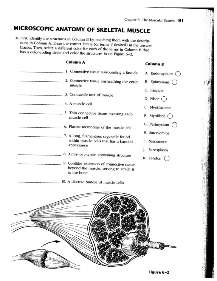
While understanding the individual muscle fiber is crucial, we must also examine how these fibers work together to form a functional muscle. Hundreds of muscle fibers are bundled together, enveloped in a connective tissue sheath called the endomysium. These bundles, resembling small cables, are further gathered into larger groups, surrounded by another connective tissue layer, known as perimysium. Imagine these cables grouped together within a larger protective layer, forming a powerful, coordinated unit.
Finally, all these bundles are enclosed by a tough outer layer called the epimysium, providing strength and support. This complex, multi-layered structure allows for efficient force transmission and ensures coordinated contraction of the entire muscle. Think of this final layer as the overall protective covering, coordinating the action of all the bundled cables within.

Image: anatomyworksheets.com
The Power of Teamwork: Nerve-Muscle Communication
Muscles don’t simply contract on their own; they receive commands from the nervous system. A single motor neuron, like a conductor in an orchestra, controls a group of muscle fibers. This unit is called a motor unit. The motor neuron sends electrical signals, instructing the muscle fibers to contract. Picture a conductor waving his baton, directing the musicians to play. The electrical signal travels along the muscle fiber, triggering the release of calcium ions. These calcium ions then bind to proteins called troponin, located on the actin filaments. This binding causes a shift in the troponin-tropomyosin complex, revealing the myosin-binding sites on the actin filaments.
With the binding sites exposed, myosin heads can now attach to the actin filaments and initiate the pulling action, leading to muscle contraction. The intensity of the contraction depends on the number of motor units recruited. The more motor units activated, the stronger the contraction.
Beyond the Basics: Exploring Muscle Adaptations
The world of skeletal muscle is far richer than just the basic structural components. Muscles adapt based on their function. For instance, endurance athletes possess a high percentage of slow-twitch muscle fibers, which are fatigue-resistant and ideal for sustained activity. Strength athletes, on the other hand, have a higher proportion of fast-twitch fibers, capable of generating bursts of power.
These adaptations are not merely a matter of genetics. Training, nutrition, and even aging can influence the distribution and function of different muscle fiber types. Understanding these adaptations allows us to tailor training programs for specific goals, whether it’s building endurance, increasing strength, or simply maintaining a healthy level of muscle mass.
Enhancing Your Understanding: Actionable Tips and Insights
Armed with this knowledge, you can now approach your own physical activities with a deeper understanding. Understanding muscle fiber types can guide your exercise choices. For example, incorporating high-intensity sprints in your routine can stimulate fast-twitch fiber development. Training with weights can increase muscle mass and strength, while endurance activities like running or cycling can improve the efficiency of slow-twitch fibers.
Remember, proper nutrition plays a critical role in muscle growth and repair. Consuming adequate protein after a workout helps your body rebuild and strengthen muscle tissue. Proper hydration is equally important, as it helps with nutrient delivery and muscle function.
Microscopic Anatomy And Organization Of Skeletal Muscle Review Sheet 11
From Microscopic Wonders to Macro-Level Results
In the vast tapestry of the human body, skeletal muscle stands as a testament to nature’s ingenuity. By understanding the intricate world of microscopic anatomy and organization, we gain a deeper appreciation for the powerful and coordinated system that allows us to move, perform activities, and experience the world around us. This journey into the microcosm of skeletal muscle empowers us to make informed decisions about our physical health, fostering a sense of awe and respect for the complex symphony of life that unfolds within us.
What excites you most about the world of skeletal muscle? Share your insights and questions, and let’s continue this journey of discovery together!


